 | Info
Sheets |
| | | | | | | | | | | | | | | | | | | | | | | | |
 | Out-
side |
| | | | |
|
| | | | |
Result : Searchterm 'T2 Star' found in 2 terms [ ] and 3 definitions [ ] and 3 definitions [ ], (+ 6 Boolean[ ], (+ 6 Boolean[ ] results ] results
| | previous 6 - 10 (of 11) nextResult Pages : [1] [2 3] |  | |  | Searchterm 'T2 Star' was also found in the following services: | | | | |
| |  |
| |
|

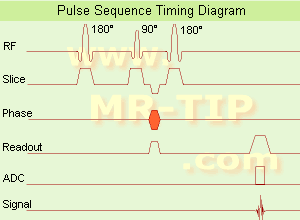
(SE) The most common pulse sequence used in MR imaging is based of the detection of a spin or Hahn echo. It uses 90° radio frequency pulses to excite the magnetization and one or more 180° pulses to refocus the spins to generate signal echoes named spin echoes (SE).
In the pulse sequence timing diagram, the simplest form of a spin echo sequence is illustrated.
The 90° excitation pulse rotates the longitudinal magnetization ( Mz) into the xy-plane and the dephasing of the transverse magnetization (Mxy) starts.
The following application of a 180° refocusing pulse (rotates the magnetization in the x-plane) generates signal echoes. The purpose of the 180° pulse is to rephase the spins, causing them to regain coherence and thereby to recover transverse magnetization, producing a spin echo.
The recovery of the z-magnetization occurs with the T1 relaxation time and typically at a much slower rate than the T2-decay, because in general T1 is greater than T2 for living tissues and is in the range of 100-2000 ms.
The SE pulse sequence was devised in the early days of NMR days by Carr and Purcell and exists now in many forms: the multi echo pulse sequence using single or multislice acquisition, the fast spin echo (FSE/TSE) pulse sequence, echo planar imaging (EPI) pulse sequence and the gradient and spin echo (GRASE) pulse sequence;; all are basically spin echo sequences.
In the simplest form of SE imaging, the pulse sequence has to be repeated as many times as the image has lines. Contrast values:
PD weighted: Short TE (20 ms) and long TR.
T1 weighted: Short TE (10-20 ms) and short TR (300-600 ms)
T2 weighted: Long TE (greater than 60 ms) and long TR (greater than 1600 ms)
With spin echo imaging no T2* occurs, caused by the 180° refocusing pulse. For this reason, spin echo sequences are more robust against e.g., susceptibility artifacts than gradient echo sequences.
See also Pulse Sequence Timing Diagram to find a description of the components.
| | | | | | | | | | | | |  Further Reading: Further Reading: | | Basics:
|
|
News & More:
| |
| |
| | | | | |
| |
|
(IR) The inversion recovery pulse sequence produces signals, which represent the longitudinal magnetization existing after the application of a 180° radio frequency pulse that rotates the magnetization Mz into the negative plane. After an inversion time (TI - time between the starting 180° pulse and the following 90° pulse), a further 90° RF pulse tilts some or all of the z-magnetization into the xy-plane, where the signal is usually rephased with a 180° pulse as in the spin echo sequence. During the initial time period, various tissues relax with their intrinsic T1 relaxation time.
In the pulse sequence timing diagram, the basic inversion recovery sequence is illustrated. The 180° inversion pulse is attached prior to the 90° excitation pulse of a spin echo acquisition.
See also the Pulse Sequence Timing Diagram. There you will find a description of the components.
The inversion recovery sequence has the advantage, that it can provide very strong contrast between tissues having different T1 relaxation times or to suppress tissues like fluid or fat.
But the disadvantage is, that the additional inversion radio frequency RF pulse makes this sequence less time efficient than the other pulse sequences.
Contrast values:
PD weighted: TE: 10-20 ms, TR: 2000 ms, TI: 1800 ms
T1 weighted: TE: 10-20 ms, TR: 2000 ms, TI: 400-800 ms
T2 weighted: TE: 70 ms, TR: 2000 ms, TI: 400-800 ms
See also Inversion Recovery, Short T1 Inversion Recovery, Fluid Attenuation Inversion Recovery, and Acronyms for 'Inversion Recovery Sequence' from different manufacturers. | | | | | |
• View the DATABASE results for 'Inversion Recovery Sequence' (8).
| | | | | | Further Reading: | | Basics:
|
|
News & More:
| |
| |
| | | | | |
| |
|
| | | | | |
• View the DATABASE results for 'Balanced Fast Field Echo' (3).
| | | | | | Further Reading: | News & More:
|
|
| |
| | | Searchterm 'T2 Star' was also found in the following services: | | | | |
| | |
| |
|
Knee MRI, with its high soft tissue contrast is one of the main imaging tools to depict knee joint pathology. MRI allows accurate imaging of intra-articular structures such as ligaments, cartilage, menisci, bone marrow, synovium, and adjacent soft tissue.
Knee exams require a dedicated extremity coil, providing a homogenous imaging volume and high SNR to ensure best signal coverage.
A complete knee MR examination includes for example sagittal and coronal T1 weighted, and proton density weighted pulse sequences +/- fat saturation, or STIR sequences. For high spatial resolution, maximal 4 mm thick slices with at least an in plane resolution of 0.75 mm and small gap are recommended. To depict the anterior cruciate ligament clearly, the sagittal plane has to be rotated 10 - 20° externally (parallel to the medial border of the femoral condyle). Retropatellar cartilage can bee seen for example in axial T2 weighted gradient echo sequences with Fatsat. However, the choice of the pulse sequences is depended of the diagnostic question, the used scanner, and preference of the operator.
Diagnostic quality in knee imaging is possible with field strengths ranging from 0.2 to 3T. With low field strengths more signal averages must be measured, resulting in increased scan times to provide equivalent quality as high field strengths.
More diagnostic information of meniscal tears and chondral defects can be obtained by direct magnetic resonance arthrography, which is done by introducing a dilute solution of gadolinium in saline (1:1000) into the joint capsule. The knee is then scanned in all three planes using T1W sequences with fat suppression. For indirect arthrography, the contrast is given i.v. and similar scans are started 20 min. after injection and exercise of the knee.
Frequent indications of MRI scans in musculoskeletal knee diseases are: e.g., meniscal degeneration and tears, ligament injuries, osteochondral fractures, osteochondritis dissecans, avascular bone necrosis and rheumatoid arthritis. See also Imaging of the Extremities and STIR. | | | | | |
• View the DATABASE results for 'Knee MRI' (4).
| | |
• View the NEWS results for 'Knee MRI' (4).
| | | | | | Further Reading: | Basics:
|
|
News & More:
| |
| |
| | | | | |
| |
|
The T1 relaxation time (also called spin lattice or longitudinal relaxation time), is a biological parameter that is used in MRIs to distinguish between tissue types. This tissue-specific time constant for protons, is a measure of the time taken to realign with the external magnetic field. The T1 constant will indicate how quickly the spinning nuclei will emit their absorbed RF into the surrounding tissue.
As the high-energy nuclei relax and realign, they emit energy which is recorded to provide information about their environment. The realignment with the magnetic field is termed longitudinal relaxation and the time in milliseconds required for a certain percentage of the tissue nuclei to realign is termed 'Time 1' or T1. Starting from zero magnetization in the z direction, the z magnetization will grow after excitation from zero to a value of about 63% of its final value in a time of T1. This is the basic of T1 weighted images.
The T1 time is a contrast determining tissue parameter. Due to the slow molecular motion of fat nuclei, longitudinal relaxation occurs rather rapidly and longitudinal magnetization is regained quickly. The net magnetic vector realigns with B0 leading to a short T1 time for fat.
Water is not as efficient as fat in T1 recovery due to the high mobility of the water molecules. Water nuclei do not give up their energy to the lattice (surrounding tissue) as quickly as fat, and therefore take longer to regain longitudinal magnetization, resulting in a long T1 time.
See also T1 Weighted Image, T1 Relaxation, T2 Weighted Image, and Magnetic Resonance Imaging MRI. | | | | | |
• View the DATABASE results for 'T1 Time' (15).
| | | | | | Further Reading: | Basics:
|
|
News & More:
| |
| |
| | | | |
| | | |
|
| |
 | Look
Ups |
| |


